Biopsy & Histopathology
What is a biopsy?
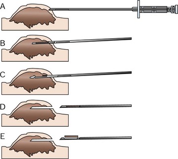
A biopsy is the surgical removal of a representative sample of tissue from a suspicious lesion. The biopsy is then processed and is examined under a microscope. The most common biopsies are:
- punch biopsy - a small, circular piece of tissue is removed using a biopsy punch,
- wedge biopsy - a small slice or chunk of tissue is removed from the tumor or mass, or
- excision biopsy - the entire mass is excised or removed.
In some cases, a biopsy it taken knowing that surgical removal of the whole tumor is not possible. In other cases, a small biopsy can be useful to plan the surgical approach or determine if other treatments may be used, thereby increasing the chances of a successful outcome. However, in many cases, particularly when it is easy to get good surgical margins (area of ‘healthy’ tissue) around the mass, it is more appropriate to remove the whole mass. Your veterinarian will give your pet a local or general anesthetic in order to take samples.
What happens to the biopsy?
After the biopsy, your veterinarian will place the tissue sample into a preservative solution and submit it to a diagnostic laboratory. In the laboratory, the process of histopathology begins.What is histopathology?
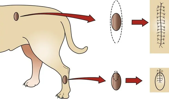
Histopathology is the examination of samples of whole tissues and is performed on a solid piece of tissue that has been collected surgically.
The piece of tissue is prepared through a process called histology by preserving, thinly slicing or sectioning, and staining the tissue sample with dyes. Once prepared, the tissue sections are examined under the microscope by a veterinary pathologist. Histopathology focuses on the architecture of the tissue and provides more information about the tissue than cytology.
With this type of laboratory examination, the accuracy of a diagnosis is usually high. The veterinary pathologist can often offer an opinion on the likely course of the disease, what is called the prognosis. If the entire mass of tissue (the whole tumor) is submitted for examination, the pathologist may also offer an opinion as to whether the tumor has been completely removed i.e., if there are wide-margins (the amount of ‘healthy’ tissue surrounding the tumor). This information helps your veterinarian to decide the best course of treatment for your pet.
What can histopathology tell me about the tumor my pet has?
It is often impossible to say what ‘lumps and bumps are just by looking at them, but with histopathology (examining cells under the microscope) the pathologist can usually assign a classification and make a diagnosis.
Cancers (malignant tumors) are usually classified by their tissue of origin and appearance.
How will I know how the cancer will behave?
As well as a diagnosis, the veterinary pathologist usually provides a prognosis, which describes the probability of local recurrence or metastasis (spread of the cancer cells to other parts of the body).
In many cases, the diagnosis with prognosis can indicate whether a cure is possible. Unfortunately, there are some cases where surgical removal and/or other treatments will only give temporary remission before recurrence or spread. The pathology results will help you and your veterinarian to plan future care for your pet.

Are there any limitations to these techniques?
The behavior of some tumors is difficult to predict. Even when a positive prognosis is made, sometimes tumors will recur or metastasize (spread to other areas). Prognoses (the likely outcomes) are based on probabilities, and nothing is 100% guaranteed.
Occasionally, particularly when it is difficult to obtain a large enough sample, a diagnosis may not be possible. Sometimes results are inconclusive, and a second biopsy is needed.