Nuclear Proptosis Surgery (Prolapse Eyeball)
What is proptosis?
Proptosis, or abnormal forward displacement of the globe out of the orbit, is a serious eye emergency that requires immediate attention to minimize discomfort and damages to the eye. With proptosis, the eyelids are trapped behind the equator of the globe and, together with profound tissue swelling in the orbit, prevent the eyeball from returning to a normal position.

What causes proptosis?
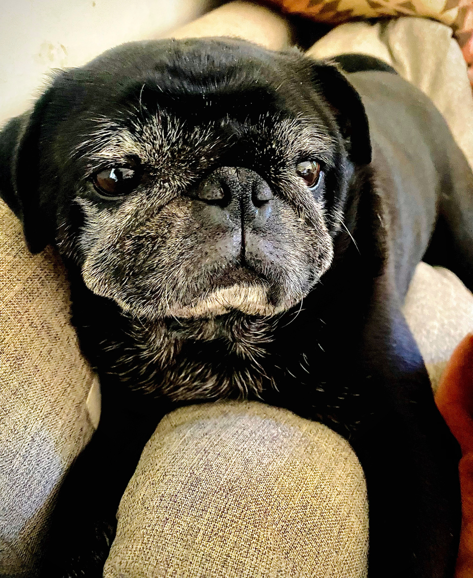
Proptosis is caused by a traumatic event such as a dog fight, blunt force injury (e.g., when a dog is hit by car), or trauma from excessive physical restraint. The latter is particularly true in brachycephalic dogs (dogs with shortened snouts); excessive pressure around the eyelids or neck (e.g., scruffing) may make the globe in these dogs protrude easily due to their shallow orbits.
How is proptosis diagnosed?
Proptosis is diagnosed based on history (trauma and acute onset) and a characteristic appearance: forward protrusion of the globe, eyelids rolling inward and being trapped behind the equator of the globe, and patient’s inability to blink the eyelids. Other signs include pain, eye swelling, haemorrhage, and strabismus (eyes do not line up in the same direction).
What are the treatment options for proptosis?
Globe replacement and temporary tarsorrhaphy (joining all of the upper and lower eyelids to completely close the eye) is generally preferred, as the globe can be removed at a later visit if needed. Cosmetic outcome is often satisfactory, and in some cases vision may be retained. Whether or not to save the globe depends on how comfortable it is to the pet and how cosmetically acceptable the pet looks to the owner.
Alternatively, enucleation should be considered as the initial treatment option if the patient has globe rupture, bleeding inside the eye, damages to the muscles or nerves around the eye or orbit/skull fractures, or if the owner is unable to provide potential long-term care.

How is globe replacement performed?
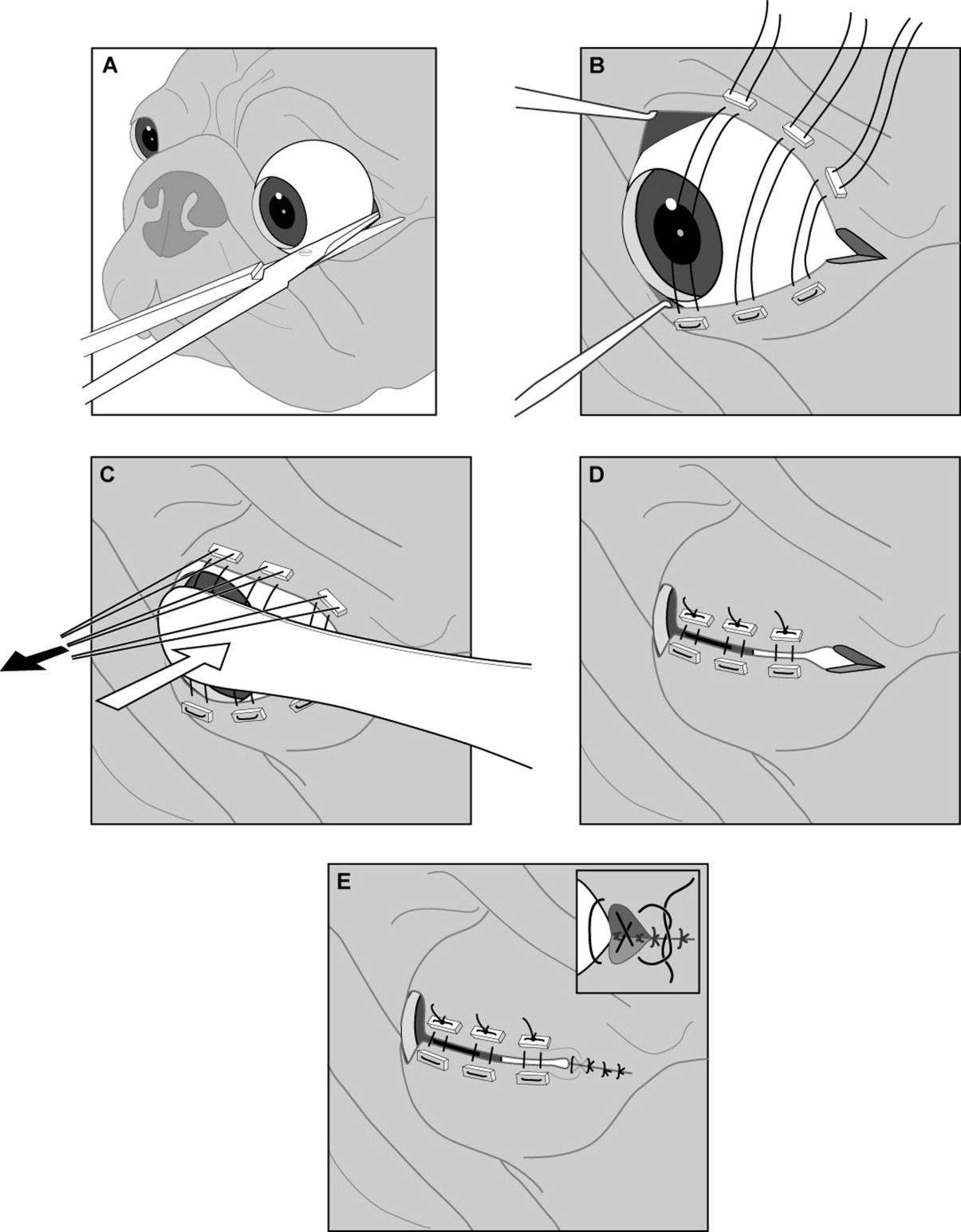
Following an ophthalmic examination, lubricant is applied to the eye (artificial tears gel or ointment) to protect the corneal surface from drying out. A thorough physical and neurologic examination is performed to ensure the patient is stable. With the patient under general anesthesia or heavy sedation, hair around the eye is clipped or trimmed, the skin around the eye is rinsed and prepared for surgery.
Globe replacement is achieved by placing sutures then bringing the eyelid margins forward by lifting the sutures and applying gentle counterpressure on the globe. This method alone is often sufficient to replace the globe. However, some patients may require a lateral canthotomy (cutting the eyelid margin to extend it) to relieve excessive tension and facilitate globe replacement. Once the globe is repositioned, sutures are placed to bring the upper and lower lids together.

What is the post-operative care?
Postoperative therapy may include antibiotics and pain medications.
Additionally, owners should look for gapping between the upper and lower eyelids, as suture material rubbing the eye surface can cause serious corneal ulceration.
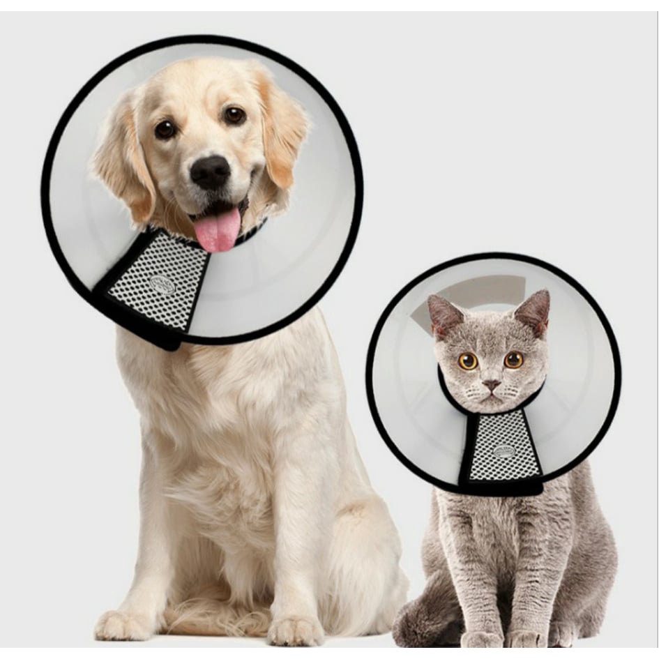
Sutures should remain in place for 2 to 3 weeks and can be removed all at once. Activity restriction and putting on Elizabethan collar are important until all sutures are removed.
What are the complications of proptosis?
Long-term complications of proptosis are common, as the traumatic displacement of the globe can damage the optic nerve, muscles around the eye, and vascular and nervous supply to the eye. Hence, owners should be aware that long-term medications may be necessary and that enucleation may be recommended in future visits if the globe is nonvisual and painful.
Keratoconjunctivitis sicca (dry eyes) and corneal ulceration are common postoperative complications; therefore, Schirmer tear test (to test whether tear production is sufficient) and fluorescein staining (to test for the presence of ulceration in the eye) may be performed after suture is removed.


