Ureteral Obstruction
What is ureteral obstruction?
Ureters are ducts carry urine from the kidney to the urinary bladder. Ureteral obstructions can be acute or chronic, unilateral or bilateral, partial or complete.
How does ureteral obstruction develop?
Congenital (present at birth) ureteral obstructions have been seen, but they are relatively rare. Acquired obstructions are more frequent. Cats are prone to ureteral obstruction because their ureters are relatively small (around 0.4mm in diameter).
Obstruction is most commonly caused by calculi (stones), which contain calcium oxalate. Stricture is also another common cause for ureteral obstruction. If your pet has previous ureteral surgery, stricture will be more likely. Obstruction can also be caused outside the ureters, for example, a space occupying lesion. Less common causes include inadvertent ureteral ligation when the pet is being spayed.
What are the clinical signs of ureteral obstruction?
Different patients have different presentations. Many have classical clinical signs such as acute pain, not being able to urinate, having blood in urine, decreased appetite and energy level, vomiting etc.
When the vet is examining your pet, if your pet’s kidneys appear to be asymmetrical i.e. one is firm and enlarged while the other is small, it is suggestive of an obstructive disease.
How is ureteral obstruction diagnosed?
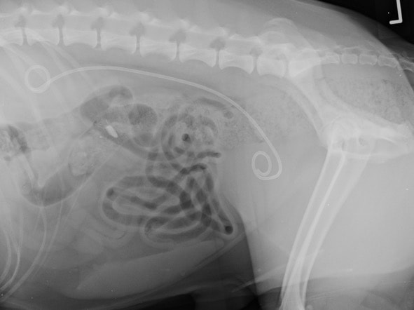
Apart from history, physical examination and clinical abnormalities, your vet will also need to do diagnostic imaging to diagnose ureteral obstruction. Abdominal radiography and ultrasonography should be use to identify the obstruction. However, not all obstructions can be easily identified, such as those caused by a stricture or small stones. It is suggested that 80% of the cases can be successfully picked up by radiographs or ultrasound. The use of both radiography and ultrasonography can increase the success rate to 90%.
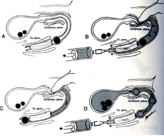
When the suspicion for ureteral obstruction is high but the obstruction cannot be identified by both imaging modalities, contrast studies or CT scan can then be considered.
Common laboratory abnormalities include low red blood cells count, high renal parameters (urea and creatinine), high potassium and high white blood cells count.
Can ureteral obstruction be treated medically?
There is no solid evidence guiding medical management of ureteral obstruction.
In theory, partially obstructing stones can pass into the lower urinary tract over time and be removed from the urinary bladder. If the patient’s renal parameters is not raised or just mildly raised and there is no evidence of obstruction on imaging, a “wait and see” approach is reasonable. It can take several weeks for the stones to migrate through the urinary tract. Regular follow-up radiographs and ultrasound examinations must be done to monitor the progression. Risks of this approach include acute obstruction and progressive deterioration of the kidney(s) and ureter(s) affected.
Fluids can be administered to facilitate the excretion of urine and the migration of small stones in the ureters. Medications that can produce or promote smooth muscles relaxation, urine excretion, anti-inflammatory effects (non-steroidal anti-inflammatory agents) will be useful. In dogs and cats, it is could be considered reasonable to monitor partially obstructive stones (or inflammatory material) for 2 – 7 days.
Surgical management of ureteral obstruction
Surgery is recommended if the obstruction is persistent or when there is infection or damage to the obstructed ureter.
Surgical methods include ureterotomy, ureteral resection and anastomosis (more commonly in dogs), ureteroneocystostomy (i.e. re-implanting the ureter to the urinary bladder), subcutaneous ureteral bypass (SUB) and renal transplantation if available. Since cats have a relatively narrow ureteral lumen, resection and anastomosis are not usually used because it may cause narrowing and re-obstruction at the anastomosis site. Subcutaneous ureteral bypass is more commonly applied in cats.
Ureteronephrectomy (surgery to remove a kidney and its ureter) is used as the last resort if the obstruction cannot be cleared or bypassed or when severe and irreversible damages or infection are present.
The veterinarian will take into account the size, location and severity of the obstruction and the renal functions of the patient to determine the best course of surgical management to be taken.
What are the potential complications?
In the “wait and see” approach, progressive ureteral obstruction, persistent stone fragments, and rupture of the ureter are the potential complications. After surgical management, it is not uncommon to have urine leaking from the ureter into the abdomen.
Recurrence of urinary stones is common in dogs and cats. In the long-run, small animals should have regular follow-up examinations to monitor for urinary tract infection and assess progression or recurrence of urinary stones, renal function, urine pH and composition.
What is the prognosis of ureteral obstruction?
Prognosis for ureteral obstruction in dogs is generally good. Prognosis for ureteroliths in cats is good if the urinary stone passes, or if surgical removal is successful before a significant loss of renal function.
Hyperkalemia (too high potassium in the blood) and acidosis (too low blood’s pH) indicate poorer prognosis. Urine leakage, renal deterioration, dehiscence or stricture after surgical intervention also indicate poor prognosis.
About half of the cats are expected to sustain chronic kidney disease and have recurrent ureteral obstruction. Therefore, patients with ureteral obstruction should be closely monitored regularly for recurrence.