(TECABO) Total Ear Canal Ablation and Bulla Osteotomy
What is a TECABO?
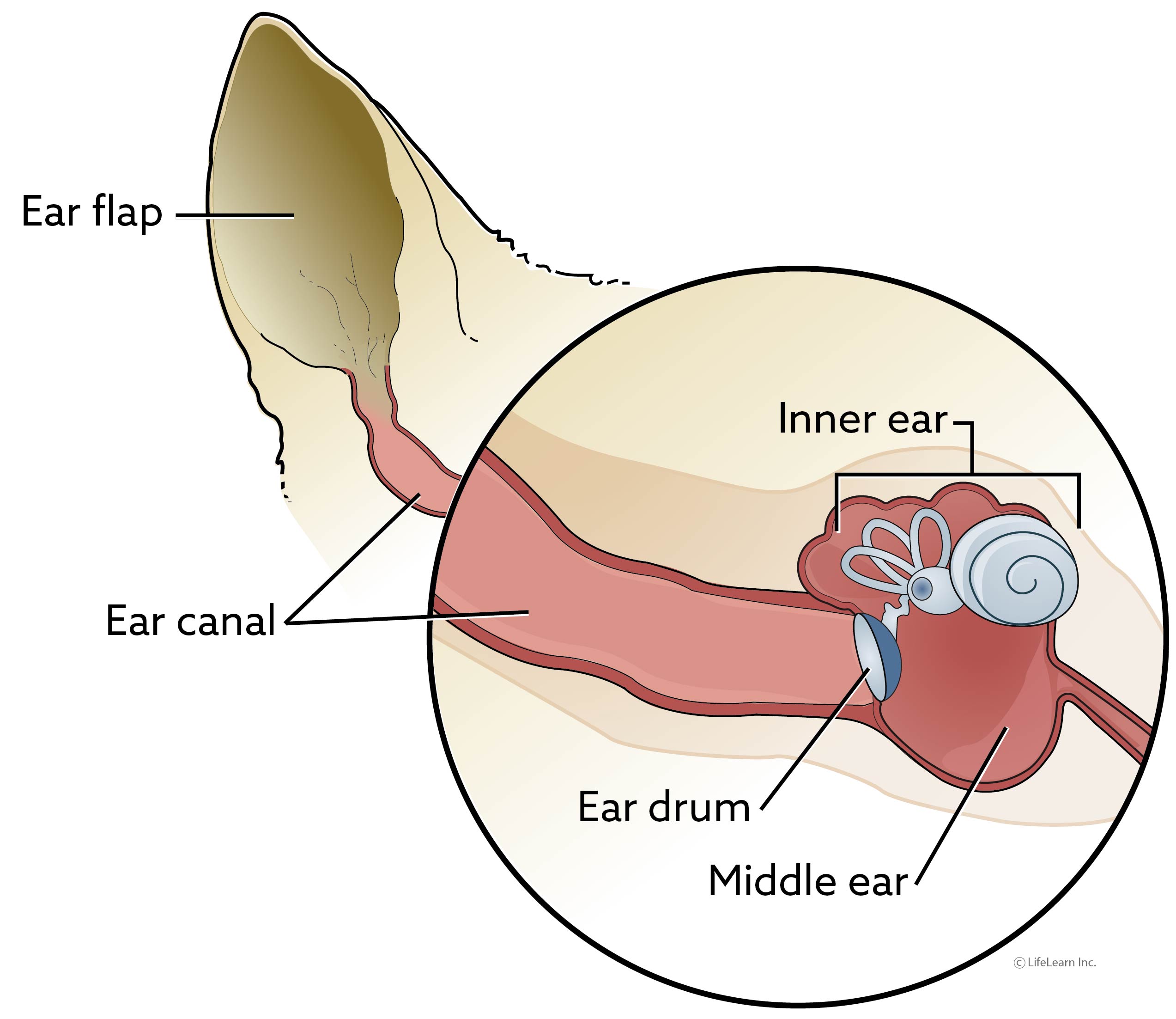
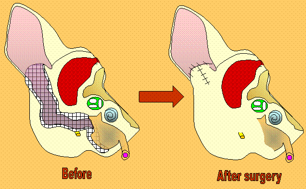
The term TECA-BO is an abbreviation for Total Ear Canal Ablation and Bulla Osteotomy. This surgery involves the complete removal of the ear canal and tympanic bulla (middle ear), leaving only the ear flap (pinna) remaining.
Why would a veterinarian recommend a TECA-BO?
A TECA-BO is primarily recommended in cases of chronic, end-stage otitis (ear infections), in which medical treatment is no longer helping the patient. In some cases, this may be due to a bacterial infection that is resistant to antibiotic treatment; removing the bacteria may be the most effective means of dealing with the infection. In many cases, longstanding infection and inflammation have led to so much scarring and mineralization of the ear canal that the ear canal has narrowed and ear cleaning is no longer effective for removing accumulated debris. In either case, a TECA-BO allows the infected, abnormal ear tissue to be removed, reducing chronic pain and inflammation and giving the pet an improved quality of life.
A TECA-BO may also be recommended in dogs or cats who have neoplastic (cancerous) growths within the ear canal. If the mass is fully confined to the ear canal, a TECA-BO will allow removal of the entire mass.
What does the TECA-BO surgery entail?
Your pet will first undergo a pre-operative assessment. During a physical exam, the veterinarian will assess the extent of your pet’s ear abnormalities and assess the function of those nerves that run adjacent to the ear canal. Pre-anesthetic blood tests will be used to evaluate your pet’s internal organ function prior to anesthesia. Finally, imaging will be performed to evaluate the ear canals and bullae (middle ear). Although X-rays can be used to image the skull and bullae, advanced imaging such as CT scan or MRI are often performed to allow more effective visualization. This pre-operative assessment will assist the surgeons in planning your pet’s surgery.
Your pet will be placed under general anesthesia for surgery. The surgeon will create a skin incision surrounding the ear, then carefully cut through the underlying tissues so that the ear canal can be removed as one intact cylinder. The ear drum and the bones of the middle ear will also be removed. This will expose the middle ear cavity, also known as the tympanic bulla. Infected material will be removed from the bulla and submitted for bacterial culture. This culture will allow identification of the infection-causing bacteria, as well as a determination of the most effective antibiotics for treatment. The bone lining the bulla will be scraped clean and the incision will be closed. An external drain may be left in place at the surgical site, in order to allow remaining fluid/material to exit the incision.
What post-operative care will be required after TECA-BO surgery?
After surgery, your pet may be sent home with a drain still in place. There may also be bandages covering the surgical site
Your pet will also go home with both pain medications and antibiotics. Antibiotics are typically continued for 2-4 weeks following surgery, depending on your pet’s overall health status and severity of the ear canal disease.
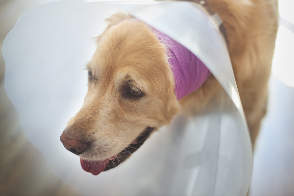
As the incision is healing, your pet will be required to wear an E-collar (cone). This will keep him from scratching at the surgical site, which could damage the incision and interfere with healing.
What complications are associated with TECA-BO surgery?
The primary risks associated with TECA-BO surgery are associated with the veins and nerves that run in the vicinity of the ear canal. Damage to the blood supply in this area may cause a partial loss of blood supply to the ear flap. If this occurs, tissue may begin to die along the edges of the ear flap and a second surgery may be required to trim away dead tissues to prevent infection. Damage to the facial nerve may result in paralysis of the affected side of the face. In many cases, this paralysis is temporary and will resolve without treatment, but in some cases the paralysis may be permanent.
Many pet owners expect their pet’s hearing to be reduced after surgery, due to the removal of the ear drum. This is definitely a possibility, but is not always the case. In many cases, the ear canal is so diseased by the time a surgery such as TECA-BO is being considered that owners notice little change in their pet’s ability to hear after surgery.
Some patient will experience chronic drainage from the incision, indicating the presence of residual infection. This drainage may be noted months to years after the original surgery. Although the drainage will often resolve temporarily with antibiotic treatment, a second surgery is often required for complete resolution.